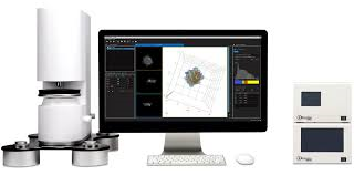
Holotomographic (3D holographic) microscopy
페이지 정보
본문
카다로그 번호 : HT-1
규격 :
브랜드 : Tomocube
비고 :
상세설명
Revolutionary holotomographic
(3D holographic) microscopy opens new era for label-free live cell imaging
Cellular analysis plays a crucial role in a wide variety of research and diagnostic activities in the life science. However, the information available to researchers and clinicians is limited by current microscopy techniques. An innovative new tool – Holotomographic microscopy – can overcome many of these limitations and open new vistas for researchers and clinicians to understand, diagnose and treat human diseases.
Holotomographic Microscopy – New era of microscopy Tomocube’s holotomography series utilize optical diffraction tomography (ODT), which enables users to quantitatively and noninvasively investigate biological cells and thin tissues. ODT reconstructs the 3D refractive index (RI) distributions of live cells and by doing so, provides structural and chemical information about the cell including dry mass, morphology, and dynamics of the cellular membrane.


TomoStudio™, Software platform for 2D/3D/4D image analysis
TomoStudio™, HT-1 operating SW, controls the system and visualizes the captured image in various ways. This flexible user interface provides fast imaging capability and 2D/3D/4D visualization of the cellular image based on 3D RI distributions of the cells and tissues. follow this link. TomoStudio™
Smart technology for holotomography
HT-1 use holotomography technology which measures the 3D and 4D refractive index (RI) tomograms. This new technology enables quantitative, label-free measurements of live, unstained cells and tissue samples. With the dynamic micromirror device (DMD), we have eliminated moving parts from light path while image-capturing and has added unprecedented stability and precision on the outstanding performance of our product. If you want to get more information about our technology, follow this link. Technology
Features and benefits
Key Features


3-D Refractive Index: No labeling
new imaging contrast


High resolution Low laser


Fast in 2D/3D High impact, Low cost
Benefits
Zero stress Label-free imaging
Intact live cell imaging Long-term image with short time interval
Time saving No sample preparation and Rapid 3D cell imaging
High quality 3D Optical resolution below 200 nm (Max. 110 nm)
Quantitative bioimaging RI enables quantitative bioimaging(local cytoplasmic concentration, dry mass)
Technical specification

Environmental requirement


