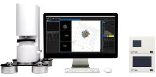
Holotomographic microscopy with 3D fluorescence
페이지 정보
본문
카다로그 번호 : HT-2
규격 :
브랜드 : Tomocube
비고 :
상세설명
HT-2 : Holotomographic microscopy with 3D fluorescence imaging capability
Research and diagnosis in the life sciences depends on the information that can be found using cellular analysis. However, current microscopy techniques limit the quantity and quality of information available to researchers and clinicians
The HT-2, a microscope that displays both 3D holograms and 3D fluorescence imaging, is able to overcome these limitations through use of new and existing technology. Its abilities can enable researchers and clinicians to break into new frontiers of understanding, diagnosing, and treating disease.
HT-2 is the world’s first microscopy combining both holotomography and 3D fluorescence imaging in one unit.
The HT-2 is an update to our HT-1 microscope. The HT-1 microscope uses holotomographic technology to measure the 3D and 4D refractive indexes using a dynamic micromirror device (DMD) to add unprecedented stability and precision to the performance of the microscope. These innovations allow for quantitative, label free imaging of cells. The HT-2 has all the capabilities of the HT-1, but combines the holotomographic imaging capabilities of the original version with the added ability to perform fluorescence microscopy.

HeLa


3D fluorescence 3D RI tomogram 3D RI tomogram 2D fluorescence
(DAPI, GFP-mitochondria) +3D fluorescence (DAPI, GFP)


Auto fluorescence – Blue Auto fluorescence – Green Auto fluorescence – Red RI tomogram
(XY cross section)
NIH-3T3


DAPI GFP – tubulin mCherry-actin RI tomogram
(XY cross section)
Features and benefits
Key Features
A three-channel LED light source
(385 nm, 470 nm, 570 nm)
Wavelengths of the LED source can be customized
Z-stack images with a motorized Z-drive (step resolution: 150 nm)
Correlative analysis in 2D, 3D and 4D with HT and fluorescence images
Upgradable: The HT-1 can be upgraded easily to the HT-2(fluorescence version) in the field
Benefits

Correlative microscopy in one instrument
HT-2 provides high-quality 3D images of both holotomography and 3D fluorescence for each sample.

Quantitative data marked with fluorescence
HT-2 provides morphological (volume, surface area, projection area, sphericity and ellipticity), chemical (dry mass, concentration) and mechanical (cell deformability) properties of cells with 3D refractive index (RI) tomogram. Moreover, fluorescence image provides information about molecular specificity.

Live cell molecular and holographic imaging with minimal stress on cells
Simultaneous measurement capability of time-lapse 3-D RI tomography and fluorescence image allows long-time tracking of specific targets in live cells. The fluorescence image provides the position of specific target organelles or structures in live cell, and consecutive measurements of time-lapse 3-D RI tomography enables the monitoring of cells and their structures with minimal stress.
Working scenarios
Scenario 1
Fast 2D fluorescence (FL) + 2D holography: enables the observation of very fast dynamics in the living cell through 2D holographic images (150 fps) after viewing the 2D FL image.


Scenario 2
Fast 2D FL + 3D HT: using a single 2D FL image, mark your target in the living cell and follow it across time using 3D HT images, without damaging the cell.


Scenario 3
3D FL + 3D HT: It is possible to obtain 3D FL and 3D HT images simultaneously, which allows researchers to investigate the 3D morphology of the cell with unprecedented imaging modalities. The quality and resolution of the 3D FL image can also be enhanced by using a deconvolution software. Learn more about our deconvolution technology here.


HT-2 fluorescence specification


